
Abdul Moiz Nadeem
Table of Contents
What is DICOM in radiology? It’s short for Digital Imaging and Communications in Medicine and is an international standard that specifies how medical images are produced, transmitted, stored, and displayed. This ensures seamless interoperability between different devices like scanners, servers, workstations, printers, PACS, etc. Before its widespread adoption, the process of image transmission was costly, time-consuming, and limited in accessibility. DICOM supports a wide range of imaging modalities, including X-rays, CT, MRI, ultrasound, nuclear medicine, and PET scans. Today, it facilitates over 98 billion medical images annually worldwide, resulting in faster, more accurate diagnoses. In the following sections, we will explore DICOM in detail, its role in radiology, components of a DICOM file type, and more.
What Is DICOM in Radiology?
DICOM is an international standard that defines how medical imaging devices like CT scanners, ultrasound machines, MRI systems, and X-rays produce, transmit, store, and display images.
It’s maintained by the DICOM Standards Committee and adopted by virtually every hospital, clinic, and imaging center in the United States and other areas.
What DICOM actually does is it wraps every medical image in a structured data package. That package includes:
- The image itself: pixel data from the scan (X-ray, CT, MRI, ultrasound)
- Patient metadata: name, ID, date of birth, medical record number
- Study information: exam type, scan date/time, body region imaged
- Technical parameters: equipment model, acquisition settings, orientation markers
This bundling ensures that an image is never separated from its clinical context. A radiologist can’t accidentally read someone else’s images. A patient’s history stays attached to their images even if files move between systems.
DICOM manages over 500 million medical images per day worldwide. Without it, each facility would be locked into one vendor’s ecosystem. Instead, healthcare IT teams can mix and match equipment, migrate to new systems, and share images securely across regions.
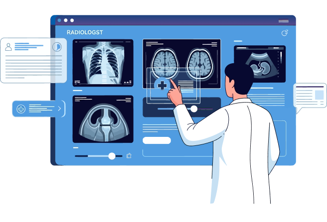
What Is a DICOM Viewer?
A DICOM viewer is specialized software that opens and displays .dcm files. Unlike a generic image viewer (which would show a blank screen on a DICOM file), a DICOM viewer:
- Parses the metadata layer to extract patient and study information
- Displays the pixel data in the correct orientation and with appropriate contrast
- Allows adjustment of window width/level (brightness/contrast) for diagnosis
- Supports multiframe display (playing ultrasound cine loops)
- Provides measurement tools (distance, angle, density)
Common DICOM viewers include:
- Standalone software: Horos (free), OsiriX, RadiAnt (Windows/Mac/Linux)
- Web-based viewers: Accessible through a portal, no installation needed
- PACS workstations: High-end diagnostic monitors with calibrated color/brightness
- Mobile apps: Allow radiologists to review studies on iPad or Android in real-time
Free viewers are fine for reviewing personal medical records. Diagnostic reading requires a calibrated monitor and specialized software to meet clinical standards.

How DICOM is Used in Radiology?
DICOM simplifies medical imaging by creating a digital standard for images and their associated data. It works as a universal language that allows different medical devices and systems to communicate.
Here’s how it’s used:
1. Image Acquisition and Storage
A radiologist orders a chest X-ray. The X-ray machine exposes film and converts it to digital data. Instead of saving as a JPEG (which would lose patient information), the machine produces a DICOM file (.dcm).
That DICOM file goes directly to the radiology department’s PACS. Think of PACS as a medical image library: infinitely searchable, automatically organized by patient, instantly accessible.
- Without DICOM: Each manufacturer’s equipment would create proprietary formats. IT teams would need separate software to read GE images versus Siemens images. Migration to new equipment would mean converting thousands of old images.
- With DICOM: Equipment from any vendor stores images in the same format. Old studies from 15 years ago open instantly alongside today’s scans.
2. Transmission and Sharing
DICOM defines how images move between systems safely. When a radiologist needs to consult a specialist at another hospital, DICOM handles the security:
- Encryption: Images transmit over TLS (standard internet security protocol)
- Authentication: Only authorized users can access images
- Audit trails: Every image view is logged, required by federal regulations (HIPAA)
A cardiologist in Boston can pull up a patient’s heart scan from a clinic in San Francisco in under 10 seconds. The image arrives with metadata intact. The cardiologist’s workstation knows it came from the San Francisco facility and logs the access.
3. Printing and Display
DICOM defines how images appear on monitors and film. This standardization prevents costly mistakes: a lesion that’s visible on one monitor should be visible on all monitors (within clinical tolerance).
Contrast and brightness are calibrated the same way across equipment. In traditional radiology, film quality varied by printer. DICOM eliminates that variability.
Why DICOM is Important in Radiology?
DICOM ensures interoperability between different imaging devices, such as MRI and CT scanners, and the PACS systems that store them.
Faster Diagnosis
A radiologist reviewing a patient’s history once had to hunt across multiple systems and vendors. Now: one query by patient ID pulls every image the patient has ever had. A comparison study that took 20 minutes now takes 30 seconds.
Seamless Communication
DICOM creates a common language for imaging devices from different manufacturers. Without this standard, each device would produce data in its own format, making it difficult to share or interpret images across platforms.
DICOM solves this problem by ensuring that all compliant equipment can communicate seamlessly, forming the backbone of modern imaging systems.
PACS Integration
DICOM integrates with PACS Medical Imaging Software. These systems are essential for storing, retrieving, and managing medical images.
Because DICOM ensures compatibility across devices, radiology teams can consolidate images from multiple modalities into a single repository, simplifying workflows and reducing errors via healthcare integration.
Specialty Workflows
DICOM enables advanced features that single-vendor systems can’t support:
- Multiplanar reconstruction (MPR): Transform a 2D CT slice into a 3D model of bones or organs
- Advanced visualization: Subtract background, highlight areas of interest, overlay measurements
- AI integration: AI models trained on DICOM datasets can flag abnormalities before a radiologist reviews the image
Standardized Data
By standardizing both image data and metadata, DICOM makes it easier for healthcare organizations to manage, store, and distribute medical images.
This eliminates the challenges of dealing with proprietary formats and enables consistent practices across entire networks.
Easy Access
With DICOM, clinicians can quickly query and retrieve images using patient identifiers or study details.
This reduces the time spent searching for specific images, streamlines reporting, and allows radiologists to focus more on interpretation rather than administrative tasks.
High-Quality Images
DICOM ensures that images retain the necessary resolution and detail required for accurate diagnoses.
Unlike compressed consumer image formats, the protocol preserves the integrity of clinical scans, allowing radiologists to rely on consistent image quality across systems.
Image Manipulation and Analysis
Beyond storage, DICOM supports advanced diagnostic functions through specialized viewers. These tools allow clinicians to adjust brightness, contrast, and zoom levels to highlight areas of concern.
Additionally, DICOM supports multiplanar reconstruction (MPR), enabling the creation of 3D views from 2D images, an essential capability in modern radiology.
Data Security
Protecting sensitive patient information is a critical priority in healthcare. DICOM includes built-in security features such as encryption, digital signatures, and access controls.
These safeguards ensure that images and associated data remain protected both during transmission and while stored in archives.
Audit Trail
The standard also supports audit trail functionality, which records data access and changes over time.
This provides transparency, enhances accountability, and helps healthcare organizations meet regulatory compliance requirements.
Long-Term Data Access
DICOM ensures that imaging data can be stored and retrieved for years after the initial scan.
This long-term accessibility is vital for follow-up care, comparative analysis, and building a comprehensive patient imaging history.

Core Components of the DICOM Standard
A DICOM file (.dcm) contains three distinct parts:
File Header (Metadata)
- Patient demographics: name, ID, date of birth, sex
- Study details: exam type (CT, X-ray, ultrasound), procedure date/time, ordering physician
- Acquisition parameters: equipment model, field of view, slice thickness, pixel spacing
- Anatomical orientation: which way is “up” in the image
Example metadata tag: (0x0010, 0x0010) = Patient Name. The tag tells DICOM readers how to interpret the value that follows.
Dataset (Image Data)
- Pixel values representing the actual image
- Single-frame (like a chest X-ray: one image)
- Multi-frame (like an ultrasound cine loop: multiple images in sequence)
Service-Object Pair (SOP) Classes
DICOM defines different types of data and what you can do with each. “CT Image Storage” is one SOP class; it means “this is a CT scan, and you can store it.” Other SOP classes include MR Image Storage, Ultrasound Image Storage, Structured Report, and Secondary Capture.
Communication Protocols
DICOM isn’t just a file format; it’s a network protocol too. Devices use DICOM’s “Store” command to send images to a PACS. They use the “Query/Retrieve” command to find and pull images. All communication happens over TCP/IP, the same protocol as email and web traffic. DICOM can layer encryption (TLS) on top of that.
What is the Image Format in DICOM?
DICOM files are in “.dcm” format, designed specifically for healthcare, and include not only the pixel data that forms the medical image but also a structured set of metadata that provides critical context.
This design allows the image to remain inseparably linked with the patient information, imaging parameters, and study details needed for clinical use.
DICOM is compatible with virtually every imaging modality in medicine, including X-ray, CT, MRI, ultrasound, nuclear medicine, PET, and digital pathology. By preserving both the image and its associated details together, DICOM ensures that medical data can be accurately interpreted, shared, and archived without risk of losing essential context.
DICOM Across Different Imaging Modalities
DICOM in Ultrasound: Handling Real-Time Data
Ultrasound differs from CT and MRI because it produces dynamic, real-time images. A single ultrasound study includes:
- Cine loops (rapid image sequences of a beating heart or moving organ)
- Doppler data (color images showing blood flow direction and velocity)
- Multi-frame images (sequences of images captured over time)
DICOM handles all of this. The standard stores frame rate information, temporal spacing between frames, and color encoding schemes. This lets a cardiologist review a patient’s heart motion years later and see exactly what the ultrasound tech saw during the original exam.
DICOM in MRI: Multi-Sequence Registration
MRI produces multiple scans of the same anatomy with different imaging parameters. A brain MRI might include:
- T1-weighted images (showing anatomy clearly)
- T2-weighted images (showing fluid/pathology)
- Diffusion-weighted imaging (DWI) (detecting acute stroke)
- 3D reconstructions (building a 3D model of a tumor)
All these sequences must be registered and aligned so they overlap perfectly. DICOM stores spatial information (voxel size, image orientation, position) that allows software to reconstruct a 3D volume or overlay sequences for comparison.
Without DICOM’s structured metadata, these sequences would exist as separate images with no way to know their spatial relationship.
DICOM in CT: High-Volume Serial Imaging
CT scans produce hundreds of thin slices. A single CT chest exam might have 500 slices, each 1mm thick. DICOM efficiently packages these into a single study with orientation information and reconstruction algorithms.
DICOM in X-Ray and Mammography
Digital X-rays and mammography images are smaller files than CT, but DICOM is still critical. Mammography systems use DICOM to embed:
- Breast density category (required for legal reporting)
- Compression force and thickness measurements
- CAD (computer-aided diagnosis) annotations
Various DICOM Protocols
The following protocols ensure that imaging workflows remain standardized, secure, and efficient.
- Network Image Management: Defines how images are securely transmitted from imaging devices to PACS or between healthcare facilities. It ensures that images arrive intact, complete, and associated with the correct metadata.
- Network Image Interpretation Management: Supports the sharing of interpretation data and structured reports, allowing radiologists and other clinicians to collaborate more effectively. This protocol enables results to travel with the images, improving communication between providers.
- Network Print Management: Provides the ability to print medical images in high resolution for physical records, patient consultations, or archival purposes. Although film is less common today, print management remains useful in certain workflows.
- Imaging Procedure Management: Governs the scheduling, execution, and documentation of imaging procedures, ensuring consistency in how imaging studies are requested, performed, and logged in hospital systems.
How Different Teams Access DICOM Files
With DICOM serving as a unified standard, all stakeholders in the healthcare system can interact with medical images consistently and reliably.
Radiologists On-Site
Radiologists access DICOM files directly through PACS imaging systems and diagnostic workstations. This workflow is pivotal to the radiology practice, as it allows clinicians to review and compare studies for accurate diagnosis and treatment planning.
Radiology teams may rely on either on-site PACS solutions, which keep data within the hospital’s IT infrastructure, or cloud-based PACS platforms, which provide scalable access across multiple locations and support remote reading.
Referring Physicians and Specialists
Outside of the radiology department, referring physicians and specialists often need to access imaging results to guide treatment decisions. DICOM files are typically delivered in a simplified viewing format through electronic health records (EHR) systems, web viewers, or shared PACS access.
This integration ensures that imaging data is readily available to surgeons, oncologists, cardiologists, and other providers who rely on radiology insights for patient care.
Patients
Patients most often access their DICOM files through patient portals provided by hospitals or imaging centers, or through physical media such as CDs, DVDs, or USB drives.
This access gives patients more control over their health information and enables them to share their medical images with new providers, specialists, or second-opinion services. Increasingly, healthcare organizations are adopting secure, cloud-based portals that allow patients to download and share DICOM studies digitally, reducing the reliance on outdated physical media.
DICOM File Format: Structure and Storage
- JPEG: Just pixels. No information about who the patient is, when the image was taken, or how to display it.
- DICOM: Pixels + patient ID + patient name + exam type + exam date + anatomical orientation + technical parameters + equipment information.
This is why you can’t open a DICOM file with a standard image viewer. A DICOM viewer must understand the metadata layer and the pixel data layer.
File Extension and Storage
The standard file extension is .dcm, but not all DICOM files have extensions. When patients receive DICOM files on a CD, the files often have no extension (just a name like “IM000001”). This is part of the DICOM standard for media archival.
DICOM files are typically large. A CT scan might be 50–200 MB. An MRI study might be 500 MB to 1 GB. These are stored in PACS databases, not local hard drives (though patients who download studies do get them on local storage).
Compression in DICOM
DICOM supports lossless compression (JPEG-LS, RLE) and lossy compression (JPEG, MPEG). Lossy compression reduces file size but slightly reduces image quality, acceptable for some uses (follow-up imaging, archival) but not for diagnostic reading of abnormalities.
The choice of compression is stored in the DICOM metadata, so any viewer knows how to decompress the image.
DICOM vs. PACS: What’s the Difference?
- DICOM is a file format and network protocol. It defines how images are structured, encoded, transmitted, and displayed. Think of it as the grammar and vocabulary.
- PACS (Picture Archiving and Communication System) is software that uses DICOM to manage a hospital’s entire imaging inventory. PACS handles:
- Storage of images on servers
- Retrieval by patient ID or study date
- User authentication and access control
- Workflow routing (which images go to which radiologist)
- Archival and backup
- Integration with the EHR
You can’t have a modern PACS without DICOM. But DICOM exists independently of any particular PACS; it’s a standard that many vendors implement.
The Future of Medical Imaging
DICOM’s structured metadata makes it ideal for AI. Machine learning models need:
- Consistent data format (DICOM provides that)
- Rich contextual information (patient age, body region, technical parameters)
- Standardized organization (all CT scans are organized the same way)
AI vendors build algorithms on DICOM datasets. An algorithm trained to detect lung nodules learns from thousands of DICOM CT scans, each with metadata about what was found and where.
When a new CT scan arrives as DICOM, the algorithm can process it because the metadata is standardized. The same algorithm works across different CT vendors.
Real-World AI Application
A radiologist in a rural clinic uploads a chest X-ray. An AI model pre-analyzes the DICOM file and flags three areas of concern.
The radiologist focuses on those areas first. Detection of a critical finding improves. The radiologist finishes the report 10 minutes faster. This is only possible because DICOM standardized the image format and metadata structure.

How PACScribe Uses DICOM
For organizations implementing DICOM workflows, PACS Medical Imaging Software like PACScribe demonstrates how they integrate with clinical systems. PACScribe, built by Folio3, brings DICOM image access directly with Epic, so radiologists can:
- Query DICOM studies without leaving their Epic integrated workflow
- View images alongside clinical notes
- Generate reports using AI-assisted interpretation
- Stay HIPAA compliant through encrypted transmission and audit logs
Interoperability
PACScribe supports the DICOM standard, which allows it to connect with imaging equipment from multiple vendors without compatibility issues.
CT, MRI, ultrasound, and X-ray studies are all stored and accessed in the same system, removing the need for manual conversions or multiple viewers. This ensures that every clinician is working with the same data in one unified platform.
Workflow Efficiency
PACScribe standardizes the intake and distribution of DICOM studies and eliminates the delays that often occur when retrieving or transferring imaging files between systems.
A radiologist can instantly query a patient’s DICOM record using ID or study date and pull up the complete set of images in seconds, rather than relying on physical media or siloed archives. This directly shortens report turnaround times and speeds up clinical decision-making.
AI-Enhanced Analysis
PACScribe integrates AI models that are trained on DICOM imaging datasets to detect specific abnormalities such as lung nodules, brain lesions, or bone fractures.
When a study is uploaded, the AI can pre-analyze the DICOM images, highlight areas of concern, and even generate structured findings. This assists radiologists by reducing oversight risk and enabling faster, more confident reporting.
Secure Compliance
Because DICOM imaging includes encryption and digital signature capabilities, PACScribe leverages these alongside HIPAA-compliant workflows to protect patient information.
All DICOM files are transmitted over secure channels, access is role-based, and audit trails log every interaction with an image.
Closing Note
DICOM is the foundation of modern medical imaging that standardizes the format and communication protocols. It ensures that radiologists, physicians, and patients alike can rely on seamless interoperability, high-quality diagnostics, and secure access to imaging data. As imaging volumes continue to grow and healthcare organizations demand greater efficiency, the importance of DICOM imaging will only deepen.
For providers looking to maximize the value of this standard, platforms like PACScribe by Folio3 Digital Health demonstrate how DICOM ultrasound can be leveraged in practice, delivering interoperability, workflow efficiency, AI-powered insights, and secure scalability. With the right solution in place, DICOM is not just a standard; it is a strategic enabler for better care, stronger collaboration, and future-ready healthcare delivery.
Frequently Asked Questions
What does DICOM stand for?
DICOM stands for Digital Imaging and Communications in Medicine.
What is a DICOM file?
A DICOM file is a standardized, comprehensive digital format for medical images like X-rays, CT scans, and MRIs, containing both the image data and metadata such as patient information, acquisition details, and diagnostic notes.
What is DICOM used for?
It is the international standard for the secure and efficient management of medical imaging data. This allows for the reliable transmission of imaging studies from a diagnostic modality (like an MRI or CT scanner) to a PACS System to a workstation for physician analysis, and to other healthcare information systems for integration with patient records.
What types of images use the DICOM standard?
DICOM is the standard for almost every type of medical image produced today, including:
- X-rays
- Computed Tomography (CT) scans
- Magnetic Resonance Imaging (MRI)
- Ultrasound
- Nuclear Medicine
- Positron Emission Tomography (PET)
- Mammography
Can a DICOM file open on a regular computer?
No. A DICOM file needs a specialized DICOM viewer software or PACS system like PACScribe by Folio3. Many of these viewers are available for free and are designed to not only display the image but also read the metadata and allow for advanced functions like measuring distances, adjusting brightness, and viewing multi-frame studies.
What is the difference between DICOM and PACS?
- DICOM is the file format and the communication rules.
- PACS (Picture Archiving and Communication System) is the centralized server system that uses the DICOM rules to manage, store, and distribute all of a hospital’s images.
DICOM is the foundation; PACS is the complete system built upon that foundation.
What Is DICOM Conformance?
DICOM Conformance refers to whether a device fully implements the DICOM standard for its use case. A device with a Conformance Statement has been tested and verified.
Without conformance, equipment might claim to be “DICOM compatible” but only support basic features, causing interoperability problems. Healthcare IT teams check Conformance Statements before purchasing to ensure seamless integration.
What is DICOM format?
A DICOM file is a special type of data container. Unlike a simple image file like a JPEG, it bundles two crucial components:
- Image Data: The actual pixel information that makes up the medical image (e.g., an X-ray, MRI, or CT scan).
- Metadata: A detailed header that contains information about the patient (name, ID, date of birth), the study (type of scan, date, time), and the equipment used. This ensures that the image is always linked to its correct clinical context.
What is the DICOM file type?
The most common file extension for a DICOM file is .dcm.
However, it’s also important to know that many DICOM files, particularly when they are on a CD or DVD for a patient, have no file extension at all. This is a common practice and is part of the DICOM standard for media storage.
What Is DICOM Ultrasound?
Ultrasound imaging using the DICOM standard. DICOM handles dynamic data (cine loops of a beating heart), color Doppler (blood flow visualization), and multi-frame sequences. This allows ultrasound studies to be stored, shared, and reviewed across systems just like CT or MRI.
What Is DICOM MRI?
MRI imaging using DICOM storage and metadata. DICOM manages multiple imaging sequences (T1, T2, DWI), multi-frame data, spatial orientation, and 3D reconstruction information. This enables seamless viewing across different vendor systems.